The ESP disc prosthesis was designed for both lumbar and cervical disc replacement. They are made of 2 titanium alloy end-plates with an elastomeric core that provides cushion and functions like a real human disc. The spikes on the outer surfaces of the end-plates improve primary fixation until the bone has developed a solid bond to the disc. The combination of a hydroxyapatite (HA) coating on top of a T40 rough surface provides an excellent bonding surface for bone ongrowth. The titanium alloy used for the end plates allows clear medical imaging and guarantees good bony fixation over time.
ESP Disc Prosthesis
ADR (Artificial Disc Replacement)


6 Degrees of Freedom
Lateral Flexion | Vertical Translation | Lateral Translation | Flexion/Extension | Anterior-Posterior Translation | Axial Rotation


- The CP-ESP and the LP-ESP discs are long-term implant devices. The disc, once implanted, is anticipated to last the lifetime of the individual.
- Tested up to 40 million cycles
- Between the 2 titanium end-plates the elastomeric parts are injection molded for controlled resistance to compression, flexion and rotation. These elastomeric parts are concentric and their fixation prohibits micro motion. The materials used for the implants have been tested for biocompatibility according to the ISO standard 10993.
- Minimally invasive anterior approach which allows reduced hospital stay and improves rehabilitation
- LP-ESP implanted since 2006 (1st version since 2004)
- CP-ESP implanted since 2012
- ESP should give a significant reduction in pain severity, re-establish spinal curvature and natural disc functions
- ESP allows quick return to normal daily activities
- Over 10 years of research and development
- 15 years of follow up (since 2004)
- 6° of freedom
- Adaptive center of rotation (unlike 1st generation ball and socket type discs with fixed center of rotation)
- No surface bearing for an increased lifetime
- Improved stability by limiting rotation and translation, which prevents overload of the posterior facet joints
- Restores elasticity and provides elastic return (unlike 1st generation ball and socket type discs which provide very little resistance and no elastic return)
- Shock absorbing
Concept of a “Silent Block” ESP®
The development of the ESP disc range originally came from Professor Roy Camille, from La Pitié Salpétrière Teaching Hospital in Paris (France). After inventing the pedicle screw which became the gold standard for Spine fusions, Pr. Roy Camille started to work on analysing the natural disc properties and designed a prosthesis to restore these.
Third Generation Disc Replacement
Both the CP-ESP and LP-ESP belong to the 3rd generation of disc replacements. Their specific monobloc design resulting from over a decade development allows them mimic the properties of natural discs. They provide movements in all directions without any friction and without any risks of debris production like other disc models.
M6 Disc Prosthesis
ADR (Artificial Disc Replacement)
The M6 artificial discs offer an innovative option compared to other artificial discs because of its unique design, which is based on the qualities of the natural disc. The M6 incorporates an artificial nucleus (made from polycarbonate urethane) and a woven fiber annulus (made from polyethylene). The M6 artificial nucleus and annulus are designed to provide the same physiologic motion characteristics of a natural disc. Extensive biomechanical testing with the M6 artificial disc has demonstrated equivalent Quality of Motion compared to a healthy disc.
Together, the M6’s artificial nucleus and annulus provide compressive capabilities and a controlled range of natural motion in all 6 degrees of freedom. This “natural” motion is designed to provide the freedom to move like a real human disc.
The M6 has two titanium outer plates with keels for anchoring the disc into the bone of the vertebral body. These outer plates are coated with a titanium plasma spray that promotes bone growth into the metal plates, providing long-term fixation and stability of the disc in the bone.


6 Degrees of Freedom
Lateral Flexion | Vertical Translation | Lateral Translation | Flexion/Extension | Anterior-Posterior Translation | Axial Rotation

- The M6-C disc and the M6-L disc are long-term implant devices. The disc, once implanted, is anticipated to last the lifetime of the individual.
- Tested up to 20 million cycles
- The M6-C Artificial Cervical Disc and the M6-L Artificial Lumbar Disc are made of the same materials. The metallic components of the device are manufactured from medical grade titanium alloy Titanium 6Al-4V. The remaining components are derived from medical grade polymers. The core and sheath are made of polycarbonate urethane (PCU), and the fiber surrounding the core is made of ultra-high molecular weight polyethylene (UHMWPE).
- Minimally invasive anterior approach which allows reduced hospital stay and improves rehabilitation
- M6-C implanted since 2005
- M6-L implanted since 2009
- M6 should give a significant reduction in pain severity, re-establish spinal curvature and natural disc functions
- M6 allows quick return to normal daily activities
- 14 years of follow up (since 2005)
- 6° of freedom
- Adaptive center of rotation (unlike 1st generation ball and socket type discs with fixed center of rotation)
- Improved stability by limiting rotation and translation, which prevents overload of the posterior facet joints
- Restores elasticity and provides elastic return (unlike 1st generation ball and socket type discs which provide very little resistance and no elastic return)
- Shock absorbing
Third Generation Disc Replacement
Both the M6-C and M6-L belong to the 3rd generation of disc replacements. Together, the M6’s artificial nucleus and annulus provide compressive capabilities and a controlled range of natural motion in all 6 degrees of freedom. This “natural” motion is designed to provide the freedom to move like a real human disc.
Before / After ADR
It took years for your disc(s) to degenerate and loose height, but it only takes a short while to regain your vertebral height through artificial disc replacement (ADR), or “spacer” placement (for fusion patients). Many patients experience almost a full inch in height gain after having the procedure.
Motion Preservation is Our Goal
While motion preservation is our goal, not everyone is a good candidate for ADR and sometimes a different procedure is the best option. Our surgeons are unbiased and highly experienced in all types of spine surgery, including endoscopic procedures and anterior / posterior fusions with minimally invasive techniques. We will recommend the best treatment and procedure for your unique case.
Use the slider to compare the before and after photos
Other Procedures at ONZ
Not everyone is a good candidate for artificial disc replacement surgery and sometimes a different procedure is the best way go. Our experienced surgeons have many tools in their toolbox to address many different types of spinal conditions. Dr. Bierstedt, Dr. Illerhaus, and Dr. Ostermann are unbiased and will recommend the best treatment for your unique case.
Some patients who want to avoid a fusion on multiple levels in their lumbar or cervical spine might be a good candidate for a hybrid procedure. With a hybrid, the patient will have a combination of ADR (artificial disc replacement) at one or more levels and a fusion at one or more levels. This hybrid solution allows the patient to still maintain some motion while decreasing the risks for adjacent level disease (sometimes called adjacent segment disease) which is common with stand alone fusions.
The Joimax TESSYS®method uses the lateral transforaminal endoscopic approach, which is, in general, a “gentle surgery.” TESSYS® can be used for all kinds of deherniation or decompression of the lumbar spine (including L5/S1). The intervertebral foramen is used for safe access to the spinal canal. In order to avoid irritation of the nerves in the foramen, the caudal part of the intervertebral foramen is widened millimeter by millimeter using special reamers. Especially, if due to location of the pathology or the age of the patient the foramen is narrow. In that case bone material has to be removed. Using special endoscopes with a working channel, herniated disc material can be removed with forceps.
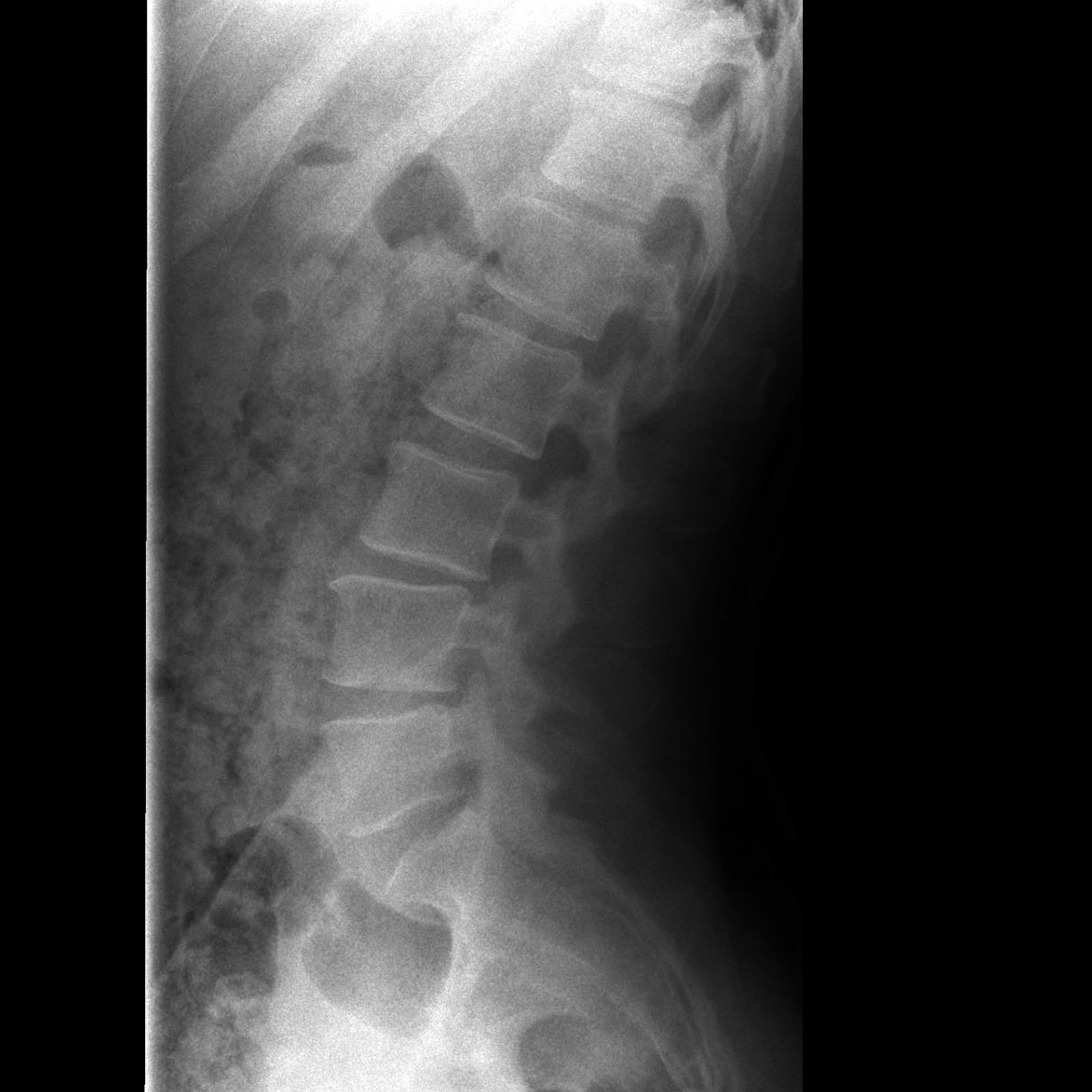
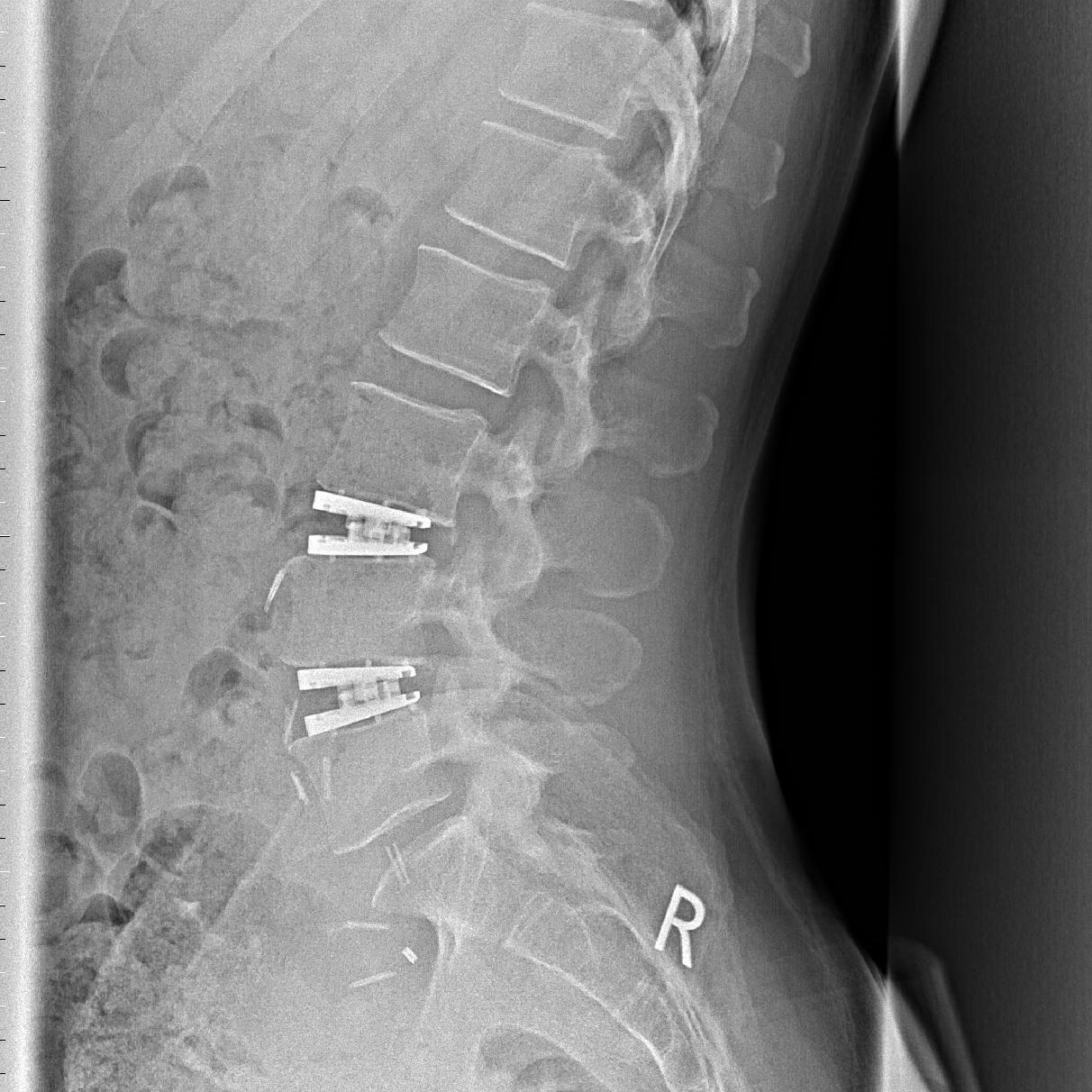
An anterior lumbar fusion is performed from the front side (through the abdomen) instead of the back. The approach is the same as with an artificial disc replacement (ADR), but instead of replacing the flesh disc with a motion preserving prosthesis, the disc is replaced with a rigid spacer or cage that is designed to fuse two vertebrae together.
Why an ALIF?
Our goal is to preserve motion at the segment when possible, but depending on the spinal condition, an anterior fusion might be the best solution for the patient. Our ultimate goal is to provide spinal stability and relieve the patient from as much pain as possible, which the ALIF allows us to accomplish.

A posterior lumbar interbody fusion is performed from the back side of the patient. As opposed to an ADR where the goal is to preserve motion, the goal for a PLIF (and ALIF) is the opposite – to stop motion. With the posterior fusion, the flesh disc is replaced with a rigid spacer or cage that is designed to fuse two vertebrae together. In addition to the spacer, rods and screws are added to correct and rebuild spinal alignment and provide the needed stability.
Why a PLIF?
Our goal is to preserve motion at the segment when possible, but depending on the spinal condition, a posterior fusion might be the best solution for the patient. Our ultimate goal is to provide spinal stability and relieve the patient from as much pain as possible, which the PLIF allows us to accomplish.

When faced with a posterior interbody fusion (PLIF), sometimes it’s possible to use a screw with a hinged joint that provides the necessary dorsal dynamic stabilization but while also preserving some mobility within the segment. These screws feature a rotation-stable joint design and Bonit coating which helps accelerate osseointegration and healing.
Why Flexible Stabilization?
Our goal is to preserve motion at the segment when possible and these flexible screws help in preserving some motion when faced with a PLIF.
Screw with Hinged Joint

Begin the Evaluation Process
Take the first step towards getting your life back and contact us today.
Copyright ©2024 all rights reserved
Visit www.onz-online.de for our full spectrum of ONZ orthopedics and neurosurgery